

Radiological
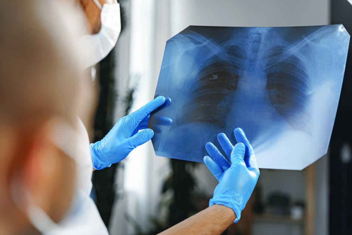
Radiology is a branch of medicine that uses various imaging technologies in the diagnosis and treatment of diseases.
Proper treatment begins with a correct diagnosis. Laboratory tests and radiologic imaging methods are crucial for accurate diagnosis.
Ankara Magnet Hospital , with this goal in mind, utilizes state-of-the-art and state-of-the-art X-ray, MRI , CT, ultrasonography, mammography , and bone densitometry (BMD) procedures performed by expert and experienced physicians. The images, meticulously evaluated using digital technology, are stored in a computer environment.
The PACS system allows our doctors to view imaging results on their screens. This system allows patients to receive rapid service and results without wasting time. When evaluated by another specialist, they can access the patient's imaging results in just a click.
- Open MRI
- Digital mammography with tomosynthesis
- Ultrasound
- Doppler ultrasound
- Bone scan
- USG-guided biopsy
- X-ray
Open MRI
High magnetic field MRI systems are also known as closed MRI systems, and although they are open on both sides, they may create a feeling of claustrophobia in some people due to their tunnel-shaped structure.
In cases where MRI examination is unavoidable for treatment, Open MRI systems are used as an alternative for diagnosis.
Ankara Magnet Hospital works in agreement with SGK and Private Health Insurance companies.Digital Mammography with Tomosynthesis
The method used to obtain serial images displays the breast's internal structure by separating it into its individual tissues. In some cases, the dense tissue within the breast can make it difficult to visualize cancerous masses.
Serial images taken with thin sections eliminate imaging errors and provide more detailed images. Because mammography devices use two-dimensional imaging, diagnosis can be challenging in some breast cancer patients. The most significant problem arises from the inability to clearly interpret overlapping structures in two-dimensional imaging.Therefore, with advancing technology, images obtained through tomosynthesis mammography are displayed in 3D, allowing for a clear view of the breast. It has been reported that using millimetric slices in tomosynthesis mammography can increase cancer diagnosis rates.
Ultrasound
Ultrasound, or ultrasonography as it is known, is a medical examination method that uses high-frequency sound waves to obtain images.
Why Is Ultrasound Used?
Ultrasound is a procedure that allows the evaluation of organs within the body and the identification of changes, developments, and problems. It is widely used to detect tumors and monitor pregnancies.
Ultrasound is also preferred during pregnancy to monitor the development of the baby, observe problems in the soft tissues and identify problems in the internal organs.
How Is an Ultrasound Performed?
This device, called ultrasound, uses sound waves at frequencies inaudible to the human ear to penetrate tissue. The real-time image generated by these sound waves can be viewed on a monitor connected to the device.
An ultrasound device consists of two separate components. The first is the probe, which comes into contact with the human body and is controlled manually. The other is the image processing unit, which converts the sound waves sent by the probe into visible information.
Doppler
Doppler ultrasound, also known as Doppler ultrasound, is a medical imaging technique similar to a traditional ultrasound. It uses radio waves, or sound waves, to visualize the body's internal structures. This method also involves no radiation.
Why Is Doppler Used?
Doppler uses stronger waves than ultrasound. Therefore, this method is preferred when more detailed imaging and imaging is required. Using Doppler, key areas such as the arms and legs can be examined. Key points on the neck can also be easily visualized with this method.
During the examination, blood flow is checked. This imaging method allows you to test for any blood flow problems.
Bone Scan
A bone scan is an imaging procedure performed after radioactive bone agents are administered intravenously into the body.
In areas where bone metabolism is increased or decreased, radioactive agent uptake is observed to change.
It is a method used in bone infections, fractures, sports injuries, tumors and similar diseases.
Why Is a Bone Scan Used?
Bone scans show that women begin to lose bone mass after age 40, and this rate increases with menopause. Women lose an average of 15% of their bone mass within the first ten years of menopause, while men lose 20-30% throughout their lives. As the rate of bone mass loss increases, the risk of fractures increases. Bone density measurements can both calculate osteoporosis and fracture risk, and facilitate post-treatment monitoring. Osteoporosis, one of the conditions that most affects women after menopause, is the thinning of the bones resulting from the loss of calcium, which increases the risk of fractures. Therefore, it is crucial to have a bone scan , perform the necessary tests and analyses, and have them monitored and evaluated by a doctor.Who Should Have Bone Density Measurement?
- If you smoke or have a personal or family history of hip fractures,
- If you are a man with diseases that accompany bone loss,
- Type 1 (insulin-dependent) diabetics, or those with a family history of osteoporosis,
- Those who show excessive collagen increases in urine tests and who have undergone high-dose bone content changes,
- Those who have fractures in their spine or other signs of osteoporosis,
- Women at high risk for osteoporosis in the premenopausal period ,
- Women who have gone through surgical menopause (both ovaries removed),
- Women with postmenopausal risk factors,
- Thin and tall post-menopausal women,
- Those with a family history of osteoporosis,
- For those with spinal fractures,
- Height less than 160 cm and weight less than 50 kilograms,
- Those who have a height loss of more than 2.5 cm per year,
- Those over 65 years of age,
- Those who have a diet poor in calcium,
- In cases of fractures due to minor impacts or traumas in adulthood,
- Those who have decreased bone density (osteopenia) detected in X-rays,
- Those who consume excessive alcohol, cigarettes and coffee,
- If there is a contraindication to estrogen therapy,
- For those with low testosterone levels,
- Those who have to remain immobile for more than 1 month,
- For diabetics,
- Those who use drugs such as heparin, antacid corticosteroids (prednisone), dilantin and barbiturates, and thyroid hormone for a long time .
Who Should Not Have Bone Density Measurement?
- For pregnant women,
- Those who have used oral or intravenous contrast material 2 to 6 days before the examination date,
- For those who cannot lie still,
- It cannot be performed on those with advanced spinal deformation.
Ultrasound-Guided Biopsy
Ultrasound -guided biopsy is performed when tissue diagnosis is required in pathologies of organs such as liver, kidney, thyroid, breast and prostate.
A biopsy performed on suspicious tissue in the body involves removing all of the unwanted tissue. Thus, a biopsy serves both diagnostic and therapeutic purposes.
Why is Ultrasound-Guided Biopsy Used and How is It Performed?
A biopsy, performed to definitively diagnose cancer, allows for the pathological examination of tissue samples taken from various parts of the body, such as the breast, brain, and thyroid, to yield definitive results. This allows for the examination of the tissue structure and, if present, the type of tumor. The results of a biopsy are also helpful in determining the treatment of the tissue and its surroundings. A biopsy does not lead to the diagnosis, the spread of the disease, or a worsening prognosis.
Fine-needle aspiration biopsies generally do not require local anesthesia. Patients may experience mild discomfort. Pain or tenderness is generally not experienced after the biopsy.
Because core needle aspiration (B) can be painful, local anesthesia is used. After the biopsy, there may be pain in the procedure area that can last 1-2 hours or tenderness that can last 1-2 days.
X-ray
X-rays visualize the inside of the body using x-rays. It is widely used to image many tissues and bones and is a painless examination. It is very useful for diagnosing, monitoring, and treating diseases.
X-rays are taken by radiographers in hospital X-ray departments.
Radiographic examination encompasses many studies that allow the internal body to be visualized using X-ray techniques. The radiation damage caused by X-rays is low.
Why is X-ray used?
An X-ray examination utilizes high-energy X-rays. These beams enter the body and are captured by tissues of varying densities. The beams, directed at a specific area of the body and emerging from the other side, are captured by a film plate. Thus, X-ray examinations allow three-dimensional tissues and organs to be projected onto the X-ray film in two dimensions, making them visible.
Taking an X-ray is quick and painless for the patient. However, this exposure creates radiation. Therefore, in children and adolescents, X-rays are one of the first steps in the initial diagnostic workup. It is beneficial to take as few X-rays as possible. X-rays are always examined and evaluated by the treatment team, along with a radiology specialist.