


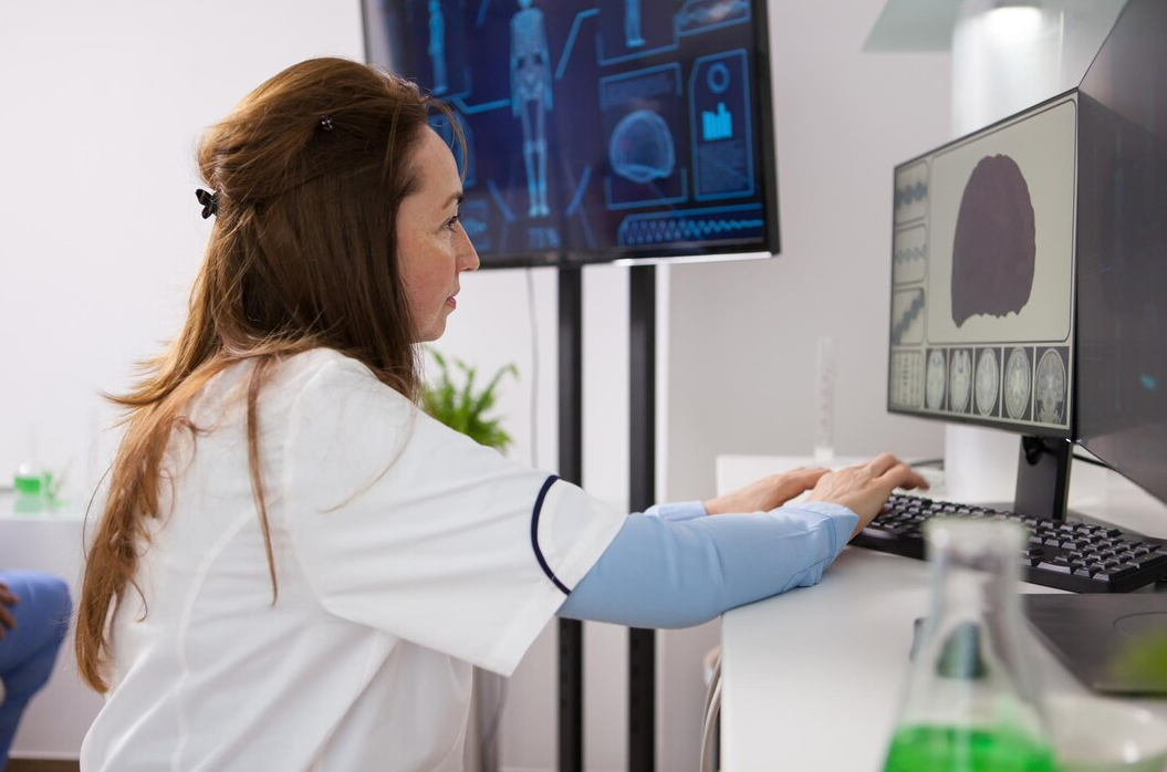
What is Virtual Angiography? Why is it Performed?
Virtual angiography is one of the most important imaging methods in modern medicine for the early diagnosis of vascular diseases . It is recommended for individuals at risk of stenosis, occlusion, or ballooning of arteries in the heart, brain, lung , kidney, abdomen, and legs . It is also a safe and comfortable option for pre-stent planning and post-stent follow-up. Early diagnosis is the most effective way to protect vascular health and improve quality of life.
Who is Virtual Angiography Recommended for?
It's recommended for people at risk of stenosis, blockage, or ballooning of arteries in the heart, brain, lung, kidney, abdomen, and legs . It's also a safe and comfortable option for pre-stent planning and post-stent follow-up. Early diagnosis is the most effective way to protect vascular health and improve quality of life.
What is Virtual Angiography?
Virtual angiography ( CT or MR angiography ) is a painless and rapid imaging method that provides a three-dimensional view of the internal structure of blood vessels . Unlike traditional angiography, this procedure involves inserting a needle through the groin; contrast material is injected into the vein, and then the vascular structure is visualized using high-resolution CT or MRI devices. This method allows for detailed examination of the heart, brain, kidney, lung, abdomen, neck, and leg vessels. This allows for the early detection of vascular stenosis, blockages, aneurysms, and congenital anomalies.
Heart and Coronary Artery Angiography (CT Coronary Angiography)
Cardiac virtual angiography is a method that provides a detailed examination of the structure of the coronary arteries that feed the heart. This examination is crucial for evaluating conditions such as coronary artery blockages , atherosclerosis (hardening of the arteries), plaque buildup, and the risk of heart attack. It provides early diagnosis in individuals with chest pain, easy fatigue, or a family history of heart disease. It is also effective in checking arterial patency after bypass surgery and the status of previously stent-implanted arteries.
Pre- and Post-Stent Virtual Angiography Checks
Pre-stent virtual angiography is performed to determine which artery to place the stent in, determine the degree of stenosis, and plan the procedure. This avoids unnecessary interventions. Post-stent virtual angiography assesses the patency and placement of the stent, and detects potential restenosis . These checks are particularly valuable in patients undergoing coronary artery stenting, as they demonstrate the stent's performance. Regular follow-up helps prevent potential complications.
Cerebral Vascular Angiography (CT/MRI Brain Angiography)
Cerebral angiography is a type of imaging that provides detailed examination of the brain's vascular network. It is particularly used to identify aneurysms (vascular ballooning) , arteriovenous malformations (AVMs), vascular occlusions, and stroke risks. It is diagnostic in patients experiencing symptoms such as dizziness, sudden vision loss, slurred speech, or temporary paralysis. It also aids in surgical planning for tumors originating from blood vessels in the brain.
Neck and Carotid (Carotid Artery) Angiography
Carotid angiography images the carotid arteries supplying the brain. Narrowing of these arteries can prevent adequate blood flow to the brain and increases the risk of stroke . Virtual angiography provides detailed images of plaque within the arteries, including their size, structure, and impact on blood flow. Early diagnosis can help patients prevent strokes through surgical or medical treatment.
Pulmonary Vascular Angiography (Pulmonary Angiography)
Pulmonary angiography is used to diagnose pulmonary embolism (lung clot) by examining the lung vessels. This procedure is vital for patients with sudden shortness of breath, chest pain, or a history of blood clots. It also provides detailed information in cases of pulmonary hypertension, congenital vascular anomalies, and vascular stenosis. This allows for the development of a rapid and effective treatment plan.
Renal and Abdominal Vessel Angiography (Renal and Abdominal Angiography)
Renal angiography visualizes the structure of the blood vessels supplying the kidneys and facilitates diagnosis of conditions such as renal artery stenosis and the causes of high blood pressure. It is also effective in diagnosing intra-abdominal vascular problems such as aortic aneurysm (a ballooning of the blood vessel), aortic dissection (a ruptured blood vessel), and liver and splenic vascular diseases. It is also frequently used for vascular mapping before organ transplantation .
Arm and Leg Vascular Angiography (Peripheral Angiography)
Peripheral angiography provides information about the condition of the blood vessels in the arms and legs. It is used to diagnose symptoms such as peripheral arterial disease, vascular occlusion, pain during walking (claudication), or slow-healing wounds. This method detects early-stage narrowing of the leg vessels, preventing serious consequences such as amputation (loss of limbs) .
Angiograms Applied to Other Organs and Surgical Planning
Virtual Angiography is used not only for disease diagnosis but also for surgical planning, identifying tumor-feeding vessels, and organ transplant preparation. It facilitates treatment planning by clarifying the vascular relationship between the disease and the brain, particularly in cases such as liver tumors, uterine fibroids, and brain tumors.
Social Security and Private Insurance Coverage
The Social Security Institution (SGK) generally covers virtual angiography procedures for diagnostic medical reasons. For patients requiring evaluation of the heart, brain, and kidney vessels, the procedure can be performed under SGK coverage, especially with a report. Private health insurance policies often cover CT and MRI angiography procedures. However, some insurance companies may charge additional fees depending on the contrast agent used or the examination center. Therefore, it is recommended to obtain approval from the insurance company before the procedure.
Frequently Asked Questions
How many minutes does virtual angiography take?
It takes approximately 10–20 minutes to complete. Including preparation and contrast medium administration, the total time does not exceed 30 minutes.
Is virtual angiography harmful?
The amount of radiation is low , and CT angiography performed with modern equipment is safe. Contrast material is used with caution in patients with kidney problems.
What is the difference between virtual angiography and classical angiography?
Virtual angiography is an imaging procedure performed without the insertion of a needle or catheter; traditional angiography involves inserting an intravenous catheter. Virtual angiography is for diagnostic purposes only, not for therapeutic purposes.
Which doctor requests virtual angiography?
Usually, a cardiologist, neurologist, cardiovascular surgeon or radiologist requests virtual angiography.
When will the results come out?
CT or MRI virtual angiography results are usually reported on the same day.
