


What is Electromyography?
Electromyography (EMG) is a medical examination that aims to evaluate nerve or muscle-related pathologies by measuring electrical activity between nerve and muscle tissues. Normally, in a healthy individual, neural impulses initiated from the brain are transmitted along nerve tissues via electrical activity, initiating a contraction process in the target tissue, the muscle fibers, through an electrical stimulus. As a result, a person can use any muscle group with the desired strength and duration.
EMG allows the measurement of electrical activity in a muscle group and the associated nerve tissue. It allows the identification of various electrical waves. These waves, received through electrical impulses from the nerve cell, are measured and projected onto a screen. Properties such as the frequency, intensity, and structure of these waves provide important information for the evaluation of various diseases affecting the muscular and nervous systems.
Why is Electromyography Performed?
An EMG (electromyography) test is used to detect and evaluate conditions affecting nerve or muscle tissue. This test is helpful in diagnosing and monitoring diseases related to the muscular and nervous systems. For patients with the following clinical symptoms, an EMG may be scheduled for further evaluation:
- The presence of twitching or intermittent involuntary contractions in the muscles
- Numbness and tingling sensation in certain areas
- Muscle weakness or difficulty in muscle movements
- Having muscle pain or frequent muscle cramps
- Feeling of chronic pain in the limbs
These symptoms may indicate a potential problem with the nervous or muscular systems. An EMG is used to identify the underlying cause of these symptoms and diagnose the disease. The test is an important tool for evaluating and diagnosing conditions such as nerve damage, muscle disorders, and nerve compression. An EMG can also be used to monitor disease progression and assess response to treatment. However, in all cases, evaluation by a physician is necessary before the test can be performed or the results interpreted.
In the EMG (Electromyography) test, the following diseases can be diagnosed in patients based on various findings:
- Muscle Diseases: EMG can detect impaired muscle function and electrical activity in cases where there is progressive destruction of muscle tissue.
- Myasthenia Gravis: In diseases such as myasthenia gravis, where there is a problem in the interaction of muscle and nerve tissue, EMG can contribute to the diagnosis by providing specific data.
- Disc Herniation: In cases where the discs at the spinal cord level cause pressure on the nerve tissues, disruption in the electrical activity of the neuromuscular tissue may occur and this can be detected by EMG.
- Carpal Tunnel Syndrome : In cases where the arm nerves are under pressure at the wrist level, the electrical activity of the nerve tissue may be negatively affected and this can be detected with EMG.
- Amyotrophic lateral sclerosis (ALS): EMG can be useful in the diagnosis and follow-up of diseases such as ALS, in which the nerve tissue responsible for stimulating the muscles in the spinal cord is destroyed.
- Polio: In cases where the spinal cord tissue responsible for muscle stimulation is destroyed, such as in polio infection, EMG can be used to determine the extent of the disease.
- Peripheral Neuropathy: EMG can be used in diagnosis when the nerve tissues surrounding the body do not function properly due to diabetes or some neurological diseases.
In all cases, these diagnoses are typically made by physicians in conjunction with various tests and clinical evaluations. EMG can play a significant role in the diagnosis and monitoring of these conditions, but all diagnostic and treatment processes are generally managed by specialist physicians.
What is Electromyography and How is it Performed?
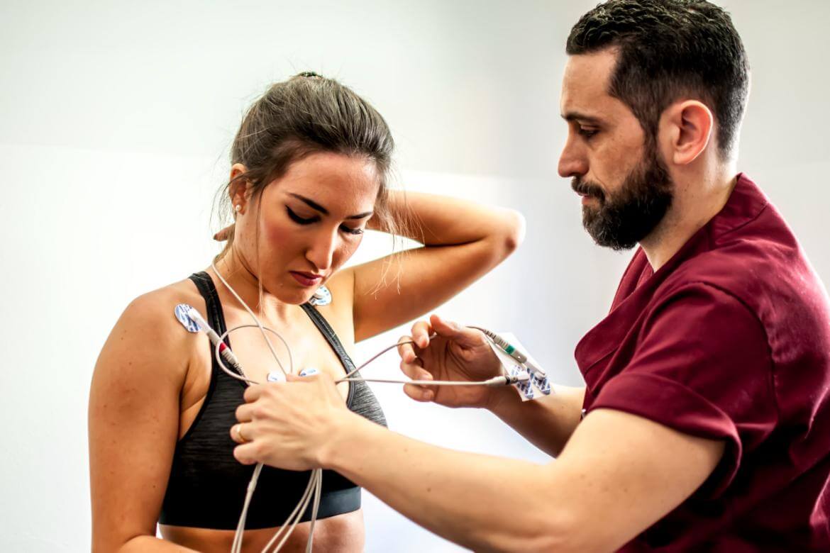
EMG (Electromyography) is a low-risk and safe medical testing method. During the procedure, a special electrode is used to measure muscle and nerve electrical activity. This electrode is placed on the targeted muscle group, and the device detects the electrical activity from the muscle and nerves. The tissue's responses are then recorded.
It's important that the area where the EMG will be performed is clean; therefore, it's recommended to shower before the procedure and avoid applying any cosmetic products to the area. Additionally, if you have electrical devices like pacemakers or bleeding disorders, you should first consult a doctor before scheduling the test.
An EMG is usually performed in an imaging laboratory by a doctor and an EMG technician. The patient lies on a chair or table. The EMG procedure typically occurs in two stages:
- Nerve activity assessment: In the first stage, nerve activity is measured with sensors placed on the skin to evaluate the functions of the nerve tissues that transmit the contraction command to the muscles.
- Evaluation of muscle electrical activity: In the second stage, after ensuring hygienic conditions for examining muscle activity and response to nerve impulses, a needle-shaped electrode is placed in the target area and electrical activity is evaluated.
An EMG typically takes 30 to 40 minutes, but this time can vary depending on the patient's condition and the area being examined. During this time, electrical activity from muscles and nerves is measured and recorded.
What should you pay attention to before an EMG examination?
Some important points to consider before an EMG (Electromyography) examination are:
- Appointment time and duration: An EMG examination is usually performed as scheduled. The procedure time can range from 30 minutes to an hour. However, sometimes previous patients' procedures may take longer, or certain circumstances may prevent the procedure from proceeding as planned. In these cases, you may have to wait. Therefore, arriving approximately half an hour before your appointment time will make the procedure smoother.
- Comfortable clothing: The patient should choose loose and comfortable clothing that can be easily taken off and put on.
- Jewelry and cosmetics: During an EMG, you may need to remove jewelry and cosmetics. It may be more appropriate to remove jewelry and avoid applying cosmetics to your skin.
- Skin hygiene: Substances such as dirt, oil, or cream on the skin can hinder the transmission of electrical signals and prolong the procedure. It may be beneficial to bathe and cleanse your skin before the procedure.
- Documentation and previous test results: It may be helpful to bring reports of your previous test results to the doctor performing the EMG. It's important to have your EMG request form and other medical reports with you.
- Notification of special conditions: Any blood-borne infectious diseases or other special health conditions should be reported to the doctor who will perform the EMG.
- Medications: The doctor should be informed about any medications used, allergies, or other important health conditions.
- Pacemaker and blood thinners: If you have a pacemaker or are taking blood thinners, it is important to inform your doctor.
- Special considerations for children: Children may lack cooperation during the procedure. Therefore, it is important to inform and reassure children before the procedure.
Finally, EMG is generally a painless and safe procedure. However, due to the needle electrodes used during the procedure, some areas may experience brief pain and, in rare cases, subcutaneous bleeding. These typically resolve quickly after the procedure.
How to interpret EMG results?
EMG (Electromyography) results are numerical data that show the electrical activity of nerve and muscle tissue in the examined area. These data are interpreted by a neurologist . Information included in the EMG report may include:
- Muscle electrical activity: Normally, muscles have a certain level of electrical activity. An EMG report evaluates the electrical activity of the muscles at rest and the activity that corresponds to contraction. In an abnormal situation, unexpected electrical activity may be observed at rest or during contraction.
- Nerve conduction velocity and conductivity : EMG can also provide information about the health of nerve tissue, such as nerve conduction velocity and the strength of the signals. This can be an indication of how quickly nerves can or cannot transmit signals.
- Identifying abnormalities: EMG results can identify abnormalities in muscles or nerves. These abnormalities can include muscle weakness, twitching (fibrillation), and nerve damage or loss.
- Diagnosis and treatment: EMG results play a critical role in disease detection and the diagnostic process. These results are crucial for doctors to determine the type of disease and develop an appropriate treatment plan.
The data contained in the EMG report is evaluated by a neurologist to determine a diagnosis of the disease or condition. Therefore, the EMG results are evaluated alongside the patient's clinical findings, complaints, and other medical test results. This evaluation determines the type, severity, and treatment plan for the disease.
How long does electromyography take?
Electromyography (EMG) is a procedure that typically takes between half an hour and an hour. However, the time can vary depending on the preparation steps and the condition of the area being examined. In some cases, factors such as the characteristics of the area being examined, the muscle groups being studied, or the patient's cooperation can lengthen or shorten the procedure. Therefore, the duration of the EMG may vary for each patient.
Is the electricity given during electromyography painful?
The electrical impulses delivered during an EMG (Electromyography) are generally low-frequency and low-intensity, so they don't cause any pain. Patients may experience a stinging sensation or mild discomfort, but they are not painful. The discomfort caused by the needle electrodes inserted into the muscles is generally minimal. In rare cases, minor bleeding may occur under the skin at the injection sites. These procedures are generally harmless, and any discomfort or pain experienced during the procedure is tolerable for most patients.
Is electromyography performed on pregnant women?
Electromyography (EMG) is a medical test that generally does not involve radiation and is generally safe for pregnant women. When considering such tests during pregnancy, doctors generally weigh the risks and benefits to determine the most appropriate option. However, EMG is generally safe to perform during pregnancy, and the risk of harm to the mother or baby is quite low. However, as with any medical test or procedure performed during pregnancy, it is important to follow your doctor's advice and recommendations. Therefore, if any medical test is necessary during pregnancy, it is important to contact your doctor and follow their recommendations.
