


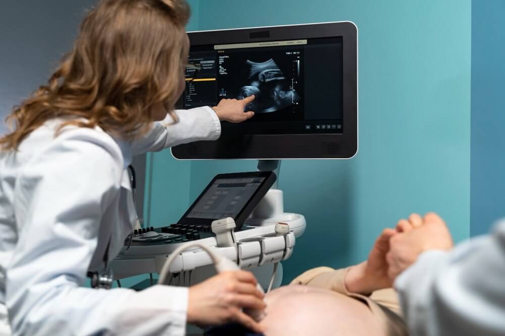
What is Abdominal Ultrasound (USG)?
An abdominal ultrasound is a painless, radiation-free diagnostic procedure that uses sound waves to image the abdominal organs (such as the liver, gallbladder, kidneys, pancreas, spleen, and bladder).
This procedure, which takes approximately 10–20 minutes, involves applying gel to the abdomen and using an ultrasound probe to evaluate the intra-abdominal organs.
Abdominal ultrasound can reveal many conditions, including fatty liver, stones, cysts, tumors, and gallbladder disease.
It is generally covered by the Social Security Institution (SGK) and private insurance companies, but prices will vary in private hospitals in 2025.
1. What is an Abdominal Ultrasound?
Abdominal ultrasound, also known as abdominal ultrasound (USG), is a safe, radiation-free diagnostic procedure that uses sound waves to image the abdominal organs. In this procedure, the ultrasound device is placed in contact with the abdominal area to examine the structure, size, and potential signs of disease of the abdominal organs. Organs such as the liver, gallbladder, kidneys, pancreas, spleen, and bladder are particularly well-known for this method.
2. How is an Abdominal Ultrasound (USG) Performed?
An abdominal ultrasound is usually performed in the radiology department. The patient is asked to undress down to the waist, and gel is applied to the abdomen. The ultrasound probe is then moved around the abdomen to visualize each organ individually. The procedure takes approximately 10-20 minutes and is painless. Results are usually reported the same day.
3. Differences Between Whole Abdominal, Upper Abdominal, and Lower Abdominal Ultrasound
a. A full-abdominal ultrasound covers both the upper and lower abdominal organs.
b. An upper abdominal ultrasound examines organs such as the liver, bile ducts, pancreas, and spleen.
c. A lower abdominal ultrasound evaluates lower abdominal organs such as the bladder, uterus, ovaries, and prostate.
The doctor determines which type of ultrasound to use based on the patient's symptoms.
4. What is an Abdominal Doppler Ultrasound?
Abdominal Doppler ultrasound is a specialized type of ultrasound that evaluates blood flow in the abdominal vessels. Color Doppler ultrasound is used to examine the vascular structure of vascular occlusions, hepatic vascular flow disturbances, or tumors.
5. What Diseases Are Seen in Abdominal Ultrasound?
The following diseases and conditions can be detected with abdominal ultrasound:
a. Liver Diseases
-
Fatty liver (hepatosteatosis)
-
Hepatitis (liver inflammation)
-
Cirrhosis (hardening of the liver)
-
Mass, cyst or tumor formations
-
Liver enlargement (hepatomegaly)
Ultrasound provides detailed information about the size, texture and surface structure of the liver.
b. Gallbladder and Bile Duct Diseases
-
Gallbladder stones (cholelithiasis)
-
Bile sludge
-
Inflammation of the gallbladder (cholecystitis)
-
Enlargement or obstruction of the bile ducts
c. Kidney Diseases
-
Kidney stones (nephrolithiasis)
-
Kidney cysts
-
Kidney enlargement or shrinkage
-
Kidney inflammation (pyelonephritis)
-
Urinary tract obstructions
c. Pancreatic Diseases
-
Inflammation of the pancreas (pancreatitis)
-
Pancreatic cyst or tumor
-
Pancreatic duct dilatations
d. Spleen Diseases
-
Spleen enlargement (splenomegaly)
-
Cysts or lesions of the spleen
-
Post-traumatic splenic rupture
e. Bladder Diseases
-
Bladder wall thickening
-
Stone or tumor formations
-
Urine accumulation (residual urine)
-
Obstruction of urine outflow
f. Uterine and Ovarian Diseases (in Women)
-
Myoma
-
Cyst
-
Polycystic ovary syndrome (PCOS)
-
Intrauterine thickening or fluid accumulation
g. Prostate and Testicular Problems (in Men)
-
Prostate enlargement
-
Prostatitis
-
Testicular cyst, varicocele or torsion
g. Intra-abdominal fluid accumulation (ascites)
Fluid accumulation in the abdominal cavity may occur due to cirrhosis, heart failure or infections.
h. Aortic Aneurysm and Vascular Diseases
Enlargement or rupture (aneurysm) of the main abdominal artery is detected.
6. Which organs are examined in an abdominal ultrasound?
The following organs are evaluated:
a. Liver
b. Gallbladder and Bile Ducts
c. Kidneys
d. Pancreas
d. Spleen
e. Bladder
f. Prostate
g. Uterus and Ovaries
g. Aorta and Vena Cava
h. Intra-abdominal fluid, lymph nodes
7. Preparation Before Abdominal Ultrasound
a. An 8-hour fast is required for an upper abdominal ultrasound.
b. A full bladder is required for a lower abdominal ultrasound.
8. Things to Consider After Abdominal Ultrasound
It does not require any special maintenance.
9. Advantages and Disadvantages of Abdominal Ultrasound
Advantages:
-
Radiation-free
-
It is painless
-
Suitable for pregnant women
-
The result is instantaneous
Disadvantages:
-
Appearance may decrease in obese people
-
The image may be reduced in gaseous bowels.
10. Alternative Imaging Methods to Abdominal Ultrasound
-
MRI
-
BT
11. Areas of Use of Abdominal Ultrasound
Abdominal pain, bloating, urinary problems, jaundice, nausea, trauma, routine screening.
12. Do Social Security and Private Insurance Cover Abdominal Ultrasound?
Generally, it is paid, there may be a difference in private hospitals.
13. In which department is abdominal ultrasound performed?
It is performed in the radiology department.
14. Who Cannot Have Abdominal Ultrasound?
It may be postponed in those with open wounds or burns.
15. What is Seen in an Abdominal Ultrasound?
Stones, cysts, tumors, fluid accumulation and vascular structures are displayed.
16. Where to Get an Abdominal Ultrasound in Ankara?
In public and private hospitals and imaging centers.
Ankara Magnet Hospital offers same-day appointments.
17. Abdominal Ultrasound 2025 Current Prices
It varies depending on the full abdomen, lower abdomen and doppler type.
18. Same-Day Abdominal Ultrasound Imaging and Appointment at Magnet Hospital
You can make an appointment online or via the hotline at 0312 212 80 80.
19. Frequently Asked Questions
a. What is Abdominal USG (Abdominal Ultrasound)?
An abdominal ultrasound is a painless, radiation-free imaging procedure that uses sound waves to visualize the internal organs of the abdomen (such as the liver, gallbladder, kidneys, spleen, pancreas, and bladder).
It is often requested for abdominal pain, bloating, or stomach or urinary problems.
b. What is checked in abdominal USG?
The structure and function of organs such as the liver, gallbladder, kidneys, spleen, pancreas, bladder, uterus, ovaries, and prostate are examined.
Intra-abdominal vessels and fluid accumulation are also evaluated.
c. How many minutes does a full abdominal USG take?
On average 10–20 minutes.
c. Why is a full abdominal USG requested?
Abdominal pain, nausea, urinary problems, elevated liver enzymes, gallstones, suspicion of kidney stones, routine check-up.
d. Which is better, ultrasound or MRI?
Ultrasound is safe, MRI is more detailed.
e. Do you remove clothes during an ultrasound?
No, only the abdomen is opened.
f. How long does a full abdominal USG take?
10–20 minutes.
g. Is inflammation seen on ultrasound?
Yes, organ inflammation can be seen indirectly.
g. Is cancer visible on ultrasound?
A suspicious mass may be seen; definitive diagnosis is MRI/CT/biopsy.
h. Why is abdominal USG requested?
Abdominal pain, indigestion, bloating, urinary problems, high enzymes, gallstones, etc.
i. Which diseases are detected in Abdominal USG?
Fatty liver, gallstones, kidney stones, pancreatitis, spleen enlargement, bladder wall thickening, ascites, etc.
i. Which department requests full abdominal USG?
Internal medicine, gastroenterology, urology, gynecology.
j. Should one be hungry or full for an abdominal ultrasound?
Upper abdomen: 8 hours of fasting.
Lower abdomen: the bladder must be full.
k. Is it done in pregnant women?
Yes, it is completely safe.
l. When will the results come out?
It is reported on the same day.
