

What is a Leg Doppler Ultrasound?
Leg Doppler ultrasound (lower extremity Doppler ultrasound) is an imaging method that uses sound waves to examine the internal structure and blood flow velocity of the leg veins. This method is particularly useful for detecting conditions such as blockages, narrowing, or clots (thrombosis) that may occur in veins and arteries. It does not involve radiation and is safe for pregnant women and the elderly.
What is Seen on a Leg Doppler Ultrasound?
Blood flow, vascular structure, and potential blockages in the leg vessels (arteries and veins) are evaluated in detail with Doppler ultrasound. Both venous and arterial Doppler ultrasounds can be performed.
1. Vascular Blockages (Thrombosis)
-
Deep vein thrombosis (DVT) clots are clearly visible.
-
It is determined whether the obstruction is complete or partial.
-
It can be understood whether the clot is new (acute) or old (chronic).
2. Varicose Veins and Venous Insufficiency
-
Vascular dilatations and valve insufficiency are demonstrated.
-
Reflux of blood is detected by color Doppler.
3. Arterial Stenosis and Occlusions
-
Plaque, narrowing, or blockage occurs in the arteries.
-
The percentage of stenosis and its effect on circulation are measured.
4. Aneurysm
-
The size and location of the ballooning in the vascular wall are determined.
5. Blood Flow Rate and Direction
-
The direction and speed of blood flow are evaluated with color codes.
6. Post-Bypass or Post-Stent Follow-up
-
The patency of the stent or bypass line is displayed.
7. Soft Tissue Findings
-
Additional findings such as edema, infection, and hematoma may be detected.
Lower Extremity (Leg) Doppler Types
Venous Doppler
It is an evaluation of the veins. It is requested in cases of DVT, venous insufficiency, and varicose veins.
Arterial Doppler
It is used to evaluate arteries. It is used to diagnose peripheral arterial disease, occlusion, and circulatory disorders.
When is Venous Doppler Ordered?
-
Sudden swelling, pain, bruising in the leg
-
Long-term bed rest, postoperative period
-
Varicose vein complaints
-
Leg edema during pregnancy
-
Leg pain after long journey
-
Recurring leg wounds
-
Pre/post varicose vein surgery check-up
-
Family history of blood clots
When is Arterial Doppler Ordered?
-
Leg pain when walking (claudication)
-
Coldness and paleness in the legs
-
Slow-healing wounds
-
Diabetic foot
-
Smoking, history of arteriosclerosis
-
Post-bypass or post-stent follow-up
-
Suspicion of post-traumatic vascular damage
Venous–Arterial Doppler Difference
|
Feature |
Venous Doppler |
Arterial Doppler |
|
Type of vein examined |
Vein |
Artery |
|
Aim |
Detection of clots, reflux, and insufficiency |
Narrowing, blockage detection |
|
The most common disease |
Deep vein thrombosis (DVT), varicose veins |
Peripheral artery disease (PAD) |
|
How to apply |
Veins are scanned along the leg |
Arteries between the knee and ankle are scanned |
|
Color Doppler advantage |
Shows clot flow and direction |
Shows flow rate and degree of stenosis |
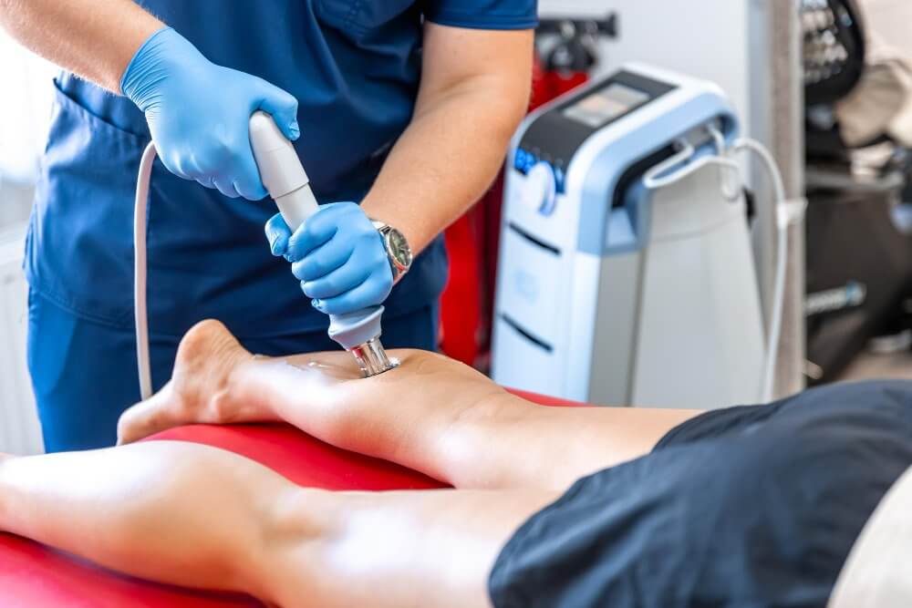
How is a Leg Doppler Ultrasound Performed?
-
The patient lies supine or prone.
-
Gel is applied to the leg and the probe is moved along the vein line.
-
It takes 15–30 minutes on average.
-
The procedure is painless and radiation-free.
-
Results are usually available the same day.
In Which Diseases Is It Taken?
-
Deep vein thrombosis
-
Peripheral artery disease
-
Arrival
-
Thrombophlebitis
-
Post-traumatic vascular damage
For which complaints is it requested?
-
Leg swelling
-
Leg pain
-
Bruising
-
Color change
-
Wound and healing problems
-
Coldness and numbness
-
Varicose vein appearance
Pre-Doppler Preparation
-
No special preparation is required.
-
Comfortable clothing is recommended.
-
If you have compression stockings, they are removed before the procedure.
Post-Doppler
-
Normal life can be returned immediately.
-
The result is available on the same day.
-
If necessary, further examination may be requested.
Disadvantages and Risks
-
Obesity, edema, or scarring may make imaging difficult.
-
It does not contain radiation and does not pose any risk of allergy.
Advantages
-
It is radiation-free.
-
It is painless and fast.
-
It is safe for pregnant women.
-
Provides early diagnosis.
-
It is cost-effective.
Alternative Imaging Methods
-
CT Angiography
-
MRI Angiography
-
IVUS (Invascular ultrasound)
Areas of Use
-
Cardiovascular diseases
-
Placental and fetal vascular flow during pregnancy
-
Vascular control after organ transplantation
-
Veins of the neck, kidney, liver, penis and abdomen
Social Security and Insurance Coverage
-
SGK covers the procedure upon doctor's request.
-
Private insurance often covers it.
-
A surcharge may apply in private hospitals.
In which department is it done?
It is performed by a radiologist in the radiology department.
To whom does it not apply?
-
Those with serious open wounds
-
Patients with leg casts
-
The procedure may be postponed in areas with intense infection.
Where to Get It Done in Ankara?
Private Magnet Hospital offers same-day appointments and fast results.
Same-Day Doppler at Magnet Hospital
-
Expert radiologists
-
Advanced color Doppler devices
-
Same day shooting and results
-
Private Hospital with SSI agreement
Frequently Asked Questions
How long does a leg Doppler scan take?
15–30 minutes.
Does it cause pain?
No.
Is it necessary to be hungry?
No need.
How many days does it take to get the results?
Same day.
Can a vascular occlusion be seen on ultrasound?
Yes.
What does the red color mean?
It indicates that blood flow is towards the device.
Will the results appear on e-Nabız?
Yes, they will be uploaded within 1–3 days.