

Liver Doppler Ultrasound
Liver Doppler ultrasound (Portal Venous Color Doppler) assesses the circulatory health of the liver by measuring blood flow, direction, and velocity in the liver vessels. This method is used in diseases such as liver cirrhosis, portal hypertension, portal vein thrombosis, hepatic vein occlusion (Budd-Chiari syndrome), liver tumors, and vascular monitoring after liver transplantation. It also significantly contributes to the diagnosis and treatment of patients with hepatitis, liver failure, coagulation disorders, and vascular anomalies.
What is Liver Doppler (Portal Venous Color Doppler) Ultrasonography?
Liver Doppler ultrasonography is a painless, radiation-free imaging method that assesses blood flow in intrahepatic vessels (specifically the portal vein, hepatic vein, and hepatic artery). This examination is crucial in diagnosing liver diseases and conditions such as cirrhosis, portal hypertension, and vascular occlusions.
How is a Liver Doppler Ultrasound Performed?
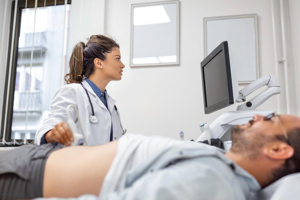
Liver Doppler ultrasound is a completely non-invasive examination, meaning it involves no intervention on the body. The patient lies supine or slightly on their right side. Gel is applied to the abdominal area, and a high-frequency ultrasound probe is used to visualize the liver's vascular structure. Color Doppler technology displays the direction and speed of blood flow within the vessels using color codes on the screen. The procedure takes approximately 15-25 minutes, and the patient experiences no pain or discomfort. Results can be evaluated immediately upon completion.
What Does a Liver Doppler Ultrasound Show?
Liver Doppler ultrasound is an advanced ultrasound technique that provides detailed images of blood flow velocity, direction, resistance, and vascular abnormalities in the liver. This examination evaluates not only liver tissue but also the function of major vessels such as the portal vein, hepatic artery, and hepatic vein.
Below, the main findings and their implications are detailed during Doppler ultrasound:
Portal Vein Flow Direction and Velocity
The portal vein is the major vessel that carries blood from the intestines to the liver.
Doppler ultrasound measures the direction (normally hepatopedal, i.e., toward the liver) and velocity of blood in this vessel.
-
If the flow is slowed, this may be a sign of portal hypertension or liver cirrhosis.
-
If blood flow is reversed (hepatofugal flow), it indicates advanced cirrhosis or severe vascular occlusion.
This measurement is one of the most valuable data about the overall health of the liver vascular system.
Portal Vein Thrombosis (Clot Occlusion) Findings
If no flow is observed within the portal vein or echogenic material is observed within the vessel lumen, this indicates the presence of a clot (thrombosis).
Doppler color mapping directly reveals areas of interrupted blood flow.
This finding is particularly important in patients with liver cirrhosis, tumors, or coagulation disorders.
Hepatic Artery Flow Resistance (Resistive Index)
The hepatic artery is the blood vessel that carries oxygen to the liver.
A Doppler blood flow measurement is made using a resistive index (RI).
-
A high RI may indicate arterial narrowing or an inflammatory process (hepatitis).
-
A low RI is an indicator of arteriovenous fistula or vascular dilatation.
These values provide direct information about the oxygenation status of the liver and circulatory health.
Expansion or Constriction of Liver Veins
Excessive hepatic vein dilatation is usually associated with causes such as portal hypertension, heart failure, or post-thrombotic reflux.
Narrowing of the veins is usually caused by vascular wall stiffness (fibrosis) or tumor compression.
Doppler ultrasound detects these changes with millimeter precision.
Blood Supply Disorder in Liver Tissue
In some cases, certain areas of the liver show inadequate blood supply (hypoperfusion).
Doppler ultrasound may show decreased or absent color flow in these areas.
This may be related to a tumor, clot, vascular occlusion, or tissue death (necrosis) following trauma.
Hepatic Vein Flow Pattern
The hepatic vein carries blood from the liver to the heart. Normally, these veins exhibit a triphasic flow pattern.
Doppler ultrasound can detect changes in this waveform:
-
Distortion of the waveform or its becoming monophasic is a sign of liver sclerosis, cirrhosis, or heart failure.
-
Loss of flow is indicative of Budd–Chiari syndrome (occlusion of the hepatic vein).
Liver Surface and Tissue Echostructure
Doppler-guided evaluation may reveal a roughened appearance, tissue heterogeneity, or nodular areas on the liver surface.
These findings are generally associated with chronic liver disease, fibrosis, or cirrhosis.
Furthermore, if a tumor or mass is suspected, Doppler ultrasound can clearly reveal blood flow differences.
Arterio-Venous Fistula or Aneurysm Findings
Abnormal connections (fistulas) or bulges (aneurysms) may form between blood vessels within the liver.
Doppler ultrasound detects very rapid, color-mixed blood flow in these areas.
This finding is often associated with liver trauma, postsurgical changes, or congenital vascular anomalies.
Increase or Decrease in Vascular Resistance
Liver vascular resistance depends on the elasticity of the vascular wall and blood flow.
-
Increased resistance usually indicates fibrosis, cirrhosis, hypertension, or an inflammatory process.
-
Decreased resistance is seen in conditions such as excessive blood flow (hyperemia) or arteriovenous shunting.
This measurement is invaluable in tracking the stage of the disease.
Vascular Structure Changes in Liver Masses
Doppler ultrasound analyzes the vascular structure of benign (e.g., hemangioma) or malignant (e.g., hepatocellular carcinoma) masses in the liver.
Increased blood flow, irregular vascular structures, or high flow velocities within the mass are generally indicative of tumor formation.
Doppler ultrasound can also monitor the survival of the tumor after treatment.
Vascular Patency After Liver Transplantation
In liver transplant patients, Doppler ultrasound is the most commonly used method in routine follow-up.
-
Portal vein flow, hepatic artery patency, and hepatic vein outflow are regularly checked with Doppler.
If any narrowing, clot or blockage is detected early, graft loss can be prevented with intervention.
Ascites (Fluid Accumulation) Around the Liver
Liver Doppler ultrasound can detect fluid accumulation (ascites) in the abdominal cavity.
This is often associated with portal hypertension or liver failure.
The amount and extent of fluid, along with any associated vascular changes, can be assessed simultaneously.
Spleen and Portal System Findings
The spleen and splenic vein are also assessed during liver Doppler.
Splenic enlargement, splenic vein dilation, or collateral vessel formation are indirect indicators of portal hypertension.
Changes Due to Medications or Toxins
Some medications, especially with long-term use, can cause increased resistance or impaired blood flow to the liver's vasculature.
Doppler ultrasound can detect these changes early and identify the risk of hepatotoxicity.
Liver Doppler ultrasound not only reveals the state of the blood vessels but also the liver's overall functional circulation, oxygenation level, and structural integrity.
The findings allow physicians to diagnose diseases such as cirrhosis, tumors, clots, vascular occlusion, or portal hypertension early and develop appropriate treatment plans.
In this respect, Doppler ultrasound is an indispensable diagnostic tool for monitoring liver diseases.
In Which Diseases is Liver Doppler Ultrasound Performed?
Liver Doppler ultrasound is a crucial method used to evaluate the liver's vascular structure and blood flow within these vessels. This examination plays a crucial role in the early diagnosis of liver diseases and in guiding treatment.
Diseases that can be detected or monitored with liver Doppler ultrasound are detailed below:
Portal Hypertension
Portal hypertension occurs due to increased pressure in the portal vein, the vessel that carries blood to the liver. Liver Doppler ultrasound aids in diagnosis by measuring the direction (hepatopedal or hepatofugal) and velocity of blood flow within the vein.
This condition, which is common in chronic liver diseases such as cirrhosis, is also a predictor of gastric and esophageal varices.
Liver Cirrhosis
Liver cirrhosis occurs when liver tissue becomes irreversibly damaged and replaced by connective tissue.
Doppler ultrasound can reveal narrowing of blood vessels and slow or reversed blood flow in cirrhosis. Findings such as increased portal vein diameter, spleen enlargement, and the presence of ascites can also be assessed with Doppler ultrasound.
Portal Vein Thrombosis (Clot Occlusion)
Portal vein thrombosis is the formation of a clot (thrombosis) in the vein that carries blood to the liver. This can lead to serious circulatory disorders.
Doppler ultrasound can directly detect the presence of a clot within the vein and, through color mapping, determine whether blood flow has stopped. This provides a significant advantage for early treatment.
Budd–Chiari Syndrome (Hepatic Vein Occlusion)
In this disease, the hepatic veins, which collect blood from the liver, become blocked. This results in increased pressure and fluid accumulation in the liver.
Doppler ultrasound reveals either no hepatic venous flow or a diminished one. This finding is highly diagnostic. It can also reveal irregularities and ascites on the liver's surface.
Liver Tumors (Hemangioma, HCC, Metastases)
Benign (hemangioma) or malignant (hepatocellular carcinoma, metastasis) tumors in the liver can cause changes in vascular structure.
Liver Doppler ultrasound can reveal the tumor's feeding vessels, blood supply pattern, and vascular compression. This provides information about the tumor type and its response to treatment.
Liver Failure
Liver failure is a serious condition in which the organ cannot maintain its functions. Doppler ultrasound can reveal findings such as decreased blood flow, vascular deformities, and decreased liver volume.
These data are used to assess the need for liver transplantation or the degree of disease progression.
Hepatitis (Acute or Chronic)
In liver inflammations such as hepatitis B, C, or autoimmune hepatitis, vascular resistance increases and changes in the hepatic artery resistive index are observed.
Doppler measurement helps determine whether inflammation is active by measuring these changes.
Post-Liver Transplant Follow-Up
In liver transplant patients, vascular patency and blood flow are regularly monitored with Doppler ultrasound.
Maintaining portal vein, hepatic artery, and hepatic vein patency is vital to preventing post-transplant complications.
Liver Vascular Anomalies
Congenital or acquired vascular abnormalities (e.g., arteriovenous fistulas, aneurysms, or malformations) can be detected with Doppler ultrasound.
These anomalies are sometimes discovered incidentally and sometimes present with signs of liver dysfunction.
Liver Metastases
If cancer that started elsewhere in the body spreads (metastasizes) to the liver, Doppler ultrasound can reveal blood vessel compression or the formation of new vascular structures.
This is an important guide in oncology treatment planning.
Thromboembolic Diseases
Clots in the liver vessels sometimes occur due to embolisms (blood clot fragments) from other vessels.
Doppler ultrasound can clearly show the interruptions in blood flow caused by these emboli.
Diseases Associated with Ascites (Abdominal Fluid) and Spleen Enlargement
In patients with abdominal fluid accumulation or splenic enlargement, Doppler imaging is used to distinguish whether the condition is vascular in origin.
Doppler findings are particularly diagnostic in cases of ascites due to portal hypertension.
Post-Thrombotic and Fibrotic Changes
Patients who have previously experienced vascular occlusion may develop fibrosis or chronic vascular narrowing.
Doppler ultrasound can detect these changes and whether blood is forming new pathways (collateral vessels).
Liver Vascular Distension Due to Heart Failure
Reflux may occur in the liver veins in right-sided heart failure.
Doppler ultrasound reveals changes in the hepatic vein waveform in these cases and is important in the differential diagnosis of cardiac-related liver failure.
Liver Damage Due to Drugs or Toxins
Some medications and toxic substances can increase vascular resistance in the liver.
Doppler imaging can reveal early changes, such as increased arterial resistance or decreased tissue perfusion, guiding treatment.
Liver Doppler ultrasonography is a rapid, reliable, and non-invasive method used in the diagnosis and monitoring of a wide range of diseases, from cirrhosis and portal hypertension to tumors and liver transplantation.
Data such as the integrity of the liver vasculature, flow direction, and velocity provide physicians with clear information about the stage of the disease, its progression, and response to treatment.
For which complaints and to whom is a Liver Doppler Ultrasound performed?
Liver Doppler ultrasound is performed on individuals experiencing symptoms such as abdominal swelling, pain, jaundice, fluid accumulation in the abdomen (ascites), weight loss, fatigue, dilated blood vessels in the skin, and elevated liver enzymes. It is also performed as a routine checkup for those with hepatitis B or C, those with liver disease due to alcohol use, those on long-term medication, and those who have undergone a liver transplant.
It can be performed safely on almost anyone, including pregnant women, and does not involve radiation.
Preparation Before Liver Doppler Ultrasound
Fasting for 6-8 hours before the procedure reduces gas formation and allows for clearer visualization of the liver vessels. Carminative medications can be used with a doctor's recommendation. Drinking plenty of water helps improve visualization of the abdominal organs.
Previous liver tests, CT scans, or MRI reports should be submitted to the physician prior to the examination.
What to Do After Liver Doppler Ultrasound
No special care is required after the procedure. The patient can return to their normal diet. However, the doctor may request further testing (CT, MRI, or biopsy) based on the Doppler results. Findings are usually reported the same day.
Disadvantages and Risks of Liver Doppler Ultrasound
Liver Doppler ultrasound, while safe, may have some technical limitations. Excessive gas, obesity, or deep-seated liver tissue may reduce image quality. However, the method has no known side effects or radiation risks. Because it does not use contrast material, allergic reactions are unlikely.
Advantages of Liver Doppler Ultrasound
The greatest advantages of this method are that it does not involve radiation, provides rapid results, and is painless. It can detect vascular occlusions early and measure blood flow velocity. It also provides information on the staging of cirrhosis, vascular patency, and changes in portal pressure after liver transplantation.
This makes it one of the most reliable non-invasive tests for guiding treatment.
Liver Doppler Ultrasonography Alternative Methods
Alternative imaging methods include CT angiography, MRI angiography, and invasive portography. However, these methods are not as safe as Doppler ultrasound because they involve contrast material and radiation.
They are generally used as a further investigation when the Doppler result is questionable.
Areas of Use for Liver Doppler Ultrasound
Liver Doppler ultrasound is a frequently used test in gastroenterology, hepatology, general surgery, radiology, and liver transplant centers. It is also used to monitor conditions such as liver vascular damage, post-tumor vascular compression, and clot formation in patients undergoing chemotherapy.
Liver Doppler Ultrasound SSI and Private Insurance Coverage
The Social Security Institution (SGK) fully covers liver Doppler ultrasound if requested by the relevant physician. Private insurance policies also generally cover this procedure at no additional cost. However, private hospitals may charge a surcharge. Institutions such as Ankara Magnet Hospital offer contracted services to SGK patients.
In Which Department Is Liver Doppler Ultrasound Performed?
This procedure is performed by the Radiology department. It is evaluated and reported by experienced radiologists. Results are usually delivered to the patient the same day.
Who Should Not Have Liver Doppler?
It may not be performed temporarily on patients with serious open wounds, surgical drains, or dressings in the abdominal area. Otherwise, it is completely safe for pregnant women, the elderly, and children.
What Does a Liver Doppler Ultrasound Show?
This examination evaluates the diameter, flow direction, velocity, and waveforms of liver vessels such as the portal vein, hepatic vein, and hepatic artery. Pathologies such as portal hypertension, thrombosis, blood flow reversal (hepatofugal flow), tumor compression, or vascular occlusion can be detected. It also provides detailed information about the progression of cirrhosis and intrahepatic blood circulation.
Where Can I Get a Liver Doppler Ultrasound in Ankara?
In Ankara, this examination is performed at university hospitals, state hospitals, and private imaging centers. In private hospitals like Magnet Hospital, it is performed by specialist radiologists. Ankara Magnet Hospital offers same-day appointments.
Liver Doppler Ultrasound Prices
Liver Doppler ultrasound costs vary depending on the location. The cost may vary depending on the center's equipment, the radiologist's experience, and the reporting time. The procedure can be performed free of charge in public hospitals under the Social Security Institution (SGK).
Ankara Magnet Hospital Liver Doppler Ultrasound - Same-Day Scan Opportunity
Ankara Magnet Hospital offers same-day liver Doppler ultrasounds without an appointment. Reports from these scans, performed by experienced radiologists, are typically prepared within an hour. Social Security Institution (SGK) agreements are available, and private insurance policies are also available. For information and an appointment, please call 0312 212 80 80.
How to Perform Liver Color Doppler?
The patient usually lies supine. Gel is applied to the abdomen, and the liver vessels (specifically the portal vein, hepatic vein, and arteries) are examined with an ultrasound probe.
Frequently Asked Questions
1. Is a liver Doppler ultrasound performed on an empty stomach?
Yes, a 6-8 hour fast is recommended.
2. Does it involve radiation?
No, it is a completely harmless method that works with sound waves.
3. When are the results available?
They are usually reported the same day.
4. Can pregnant women have it done?
Yes, it can be done on pregnant women because it is safe.
5. Is Portal Vein Color Doppler painful?
There is no pain during the procedure.
