


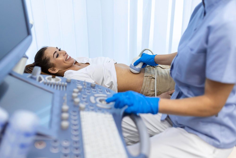
What is a Pelvic Ultrasound?
A pelvic ultrasound is a painless, radiation-free imaging method that uses sound waves to examine internal organs located in the lower abdomen (such as the uterus, ovaries, prostate, and bladder). Pelvic ultrasound provides detailed information about the reproductive organs, urinary tract, and bladder in both men and women. Therefore, it is one of the most frequently used ultrasound methods for both routine checkups and diagnosis.
Types of Pelvic Ultrasound
Pelvic ultrasound is divided into four main types according to different areas of application:
Abdominal Ultrasound: This is the classic type of pelvic examination performed on the abdomen, where the bladder must be full.
Pregnancy Ultrasound: Examines the structural features of the baby and uterus during pregnancy.
Vaginal Ultrasound ( Transvaginal USG ): It is performed vaginally to view the female reproductive organs in more detail.
Rectal Ultrasound (Transrectal USG): Used to evaluate the prostate and rectum area in men.
Each one is applied for different clinical purposes and is selected according to the doctor's guidance.
What is a Pelvic Doppler Ultrasound?
Pelvic Doppler ultrasound measures blood flow in addition to conventional ultrasound . It is used when blood flow in the ovarian, uterine, or testicular vessels needs to be assessed. Doppler imaging allows for more accurate diagnosis of conditions such as cysts, tumors, or vascular occlusions.
Pelvic Ultrasound in Women
In women, a pelvic ultrasound is used to examine the structure of the uterus, ovaries, fallopian tubes, and bladder. This method is used to evaluate ovarian cysts, fibroids , intrauterine thickening, polyps, and pregnancy monitoring. It is the most frequently requested imaging test for complaints of menstrual irregularities, abdominal pain, infertility, or vaginal bleeding.
Pelvic Ultrasound in Men
In men, a pelvic ultrasound is used to examine the bladder, prostate, seminal vesicles, and surrounding tissues. It is particularly preferred in cases of prostate enlargement , difficulty urinating, or infertility. A rectal ultrasound can be used for more detailed examination if necessary.
When Does a Doctor Order a Pelvic Ultrasound?
A doctor typically requests a pelvic ultrasound for conditions such as lower abdominal pain, urinary problems, irregular menstrual periods , infertility, abnormal bleeding, or suspected pelvic masses. It may also be requested for pre-operative or pre-treatment evaluation.
How Is a Pelvic Ultrasound Performed?
A pelvic ultrasound is usually performed with the patient lying on their back, with gel applied to their abdomen. For a vaginal or rectal ultrasound, a sterile probe is placed in the affected area using a special sheath. The procedure takes approximately 10-20 minutes and is completely painless. It does not involve radiation and is safe.
What Diseases Does Pelvic Ultrasound Indicate?
A pelvic ultrasound is an imaging procedure that provides detailed examination of the lower abdominal (pelvic) organs—the uterus, ovaries, bladder, prostate, bladder, and surrounding tissues. Therefore, it can detect a wide range of gynecological, urological, and gastrointestinal conditions.
Diseases Indicated by Pelvic Ultrasound in Women
Uterine Diseases: Uterine fibroids (myoma uteri), intrauterine polyps (endometrial polyps), uterine thickening (endometrial hyperplasia), adenomyosis (thickening of the uterine wall), intrauterine adhesions (Asherman syndrome), congenital uterine anomalies (double uterus, septum, etc.), uterine cancer (endometrial cancer) – early stage symptoms may be observed.
Ovarian Diseases: Ovarian cysts (simple, hemorrhagic, dermoid, endometrioid cysts), polycystic ovary syndrome (PCOS), ovarian torsion (rotation of the ovary around itself), ovarian tumors (benign or malignant), endometriosis (chocolate cyst).
Tube and Pelvic Infections: Fluid accumulation in the tubes (hydrosalpinx), pelvic inflammatory disease (PID), inflammation or abscess formation in the tubes.
Pregnancy-Related Conditions: Early pregnancy follow-up, ectopic pregnancy, intrauterine gestational sac and heart rate control, threatened or incomplete miscarriage, placenta and intrauterine bleeding foci.
Diseases Indicated by Pelvic Ultrasound in Men
Prostate and Bladder Diseases: Enlarged prostate (BPH), prostate inflammation (prostatitis), prostate cyst or calcification, bladder stones, bladder tumors or thickening.
Urinary Tract Problems: Incomplete emptying of the bladder (residual urine), enlargement of the urinary tract (sub-signs of hydronephrosis), urethral obstruction or stricture.
Testis and Surrounding Tissues (by Transrectal or Scrotal Pelvic USG): Testicular cyst, tumor or varicocele enlargement, epididymitis (testicular inflammation).
Conditions Common to Both Gender
Free fluid in the pelvic region (blood, inflammation, signs of infection), mass or tumor formations, abscess or infected fluid accumulation, extension of intestinal masses to the pelvis.
A pelvic ultrasound clearly shows pelvic organs such as the uterus, ovaries, prostate, and bladder. In women, it's most commonly used to detect conditions such as fibroids, cysts, endometriosis, and ectopic pregnancies, and in men, it's used to detect prostate enlargement, bladder stones, or tumors.
For Which Complaints is Pelvic Ultrasound Performed?
A doctor may order a pelvic ultrasound for symptoms such as irregular menstrual periods, groin pain, infertility, difficulty urinating, frequent urination, or swelling or pain in the lower abdomen. It is also used in routine gynecological or urological checkups.
Things to Do Before a Pelvic Ultrasound
For an abdominal pelvic ultrasound, the bladder needs to be full. Therefore, it is recommended to drink fluids 1-2 hours before the procedure. For vaginal or rectal ultrasounds, the bladder needs to be empty. It is recommended that the patient wear comfortable clothing.
What to Do After a Pelvic Ultrasound
No special care is required after the procedure. The patient can continue with their normal daily activities. Once the gel residue is removed, it leaves no lasting effects on the body.
Advantages, Disadvantages, and Risks of Pelvic Ultrasound
It's a painless, fast, and safe method. It can even be used during pregnancy. Its biggest advantages include its lack of radiation, instant results, and affordability. Ultrasound doesn't involve radiation, so the risk is low. Only brief discomfort may occur with vaginal or rectal applications. There is no risk of serious complications.
Alternative Methods
When a more detailed examination of the pelvic area is necessary, MRI ( Magnetic Resonance Imaging ) or CT (Computed Tomography) may be preferred. However, these methods are generally performed after an ultrasound if deemed necessary.
Other Uses of Pelvic Ultrasound
Pelvic ultrasound can be used not only for diagnosis but also as a guide for interventional procedures such as biopsy procedures, intrauterine device (IUD) insertion, ovarian cyst drainage or fluid aspiration.
How Long Does a Pelvic Ultrasound Take?
The average procedure time is between 10 and 20 minutes. Doppler examinations or detailed evaluations may extend this time.
Do Social Security and Private Insurance Cover Pelvic Ultrasound?
The Social Security Institution (SGK) covers pelvic ultrasounds that are deemed necessary by a doctor and performed upon referral. It is also generally covered by private insurance policies. However, private hospitals may charge a surcharge.
In Which Department Is Pelvic Ultrasound Performed?
Pelvic ultrasounds are performed by the radiology department. The patient is referred to a gynecologist, urologist, or internal medicine specialist.
Who Should Not Have a Pelvic Ultrasound?
Vaginal ultrasounds are performed in early pregnancy at the discretion of a doctor. Rectal ultrasounds are not performed during periods of active infection or after surgery in the rectal area.
What Do Pelvic Ultrasound Results Show?
The final report includes findings such as organ size and shape, the presence of masses or cysts, fluid accumulation, and vascular structure. This information supports the diagnosis and, if necessary, allows for further testing.
Where to Get a Pelvic Ultrasound in Ankara?
Pelvic ultrasounds can be performed at university hospitals, private medical centers, and institutions such as Magnet Hospital in Ankara . Same-day procedures can be performed by scheduling an appointment with the hospital's radiology department.
Pelvic Ultrasound Prices
Pelvic ultrasound prices vary depending on the type of hospital. The cost may increase if a Doppler ultrasound is added. Private hospitals often offer same-day evaluations without an appointment.
Same Day Shooting – Ankara Magnet Hospital
Ankara Magnet Hospital offers same-day evaluations for pelvic ultrasounds without an appointment. We have a partnership with the Social Security Institution (SGK), and results can be delivered to your doctor the same day.
Frequently Asked Questions
How many minutes does a pelvic ultrasound take?
The process takes approximately 10–20 minutes on average.
Is a pelvic ultrasound performed during pregnancy?
It is done safely because it does not contain radiation.
Can inflammation be seen with a pelvic ultrasound?
Yes, signs of fluid accumulation or infection in the pelvic area can be detected on ultrasound.
Do you remove clothes during an ultrasound?
In abdominal ultrasound, only the abdominal area is usually opened, and special drapes are used for vaginal or rectal applications.
Can a pelvic ultrasound be performed during menstruation?
Yes, a pelvic ultrasound can generally be performed during menstruation. However, if a vaginal ultrasound is performed, it is not recommended during heavy menstrual bleeding. An abdominal pelvic ultrasound can be performed safely during menstruation.
Can the cervix be seen in a pelvic ultrasound?
Yes, the cervix is usually clearly visualized during a pelvic ultrasound. Vaginal ultrasound, in particular, allows for a detailed assessment of the cervical length, structure, and potential lesions.
How many minutes does a pelvic ultrasound take?
The procedure takes approximately 10 to 20 minutes. If a Doppler ultrasound or detailed examination is required, the time may be extended by a few minutes.
Where is the pelvic area?
The pelvic region is the area in the lower abdomen between the navel and the thigh (groin). This region contains the uterus, ovaries, and bladder in women, and the prostate and bladder in men.
Are pelvic and groin the same thing?
They are somewhat similar areas, but not exactly the same. The groin area is located in the lower part of the pelvic region. The pelvic region is larger and includes the internal organs, while the groin is the area closer to the outside, between the thighs and abdomen.
Why is a pelvic ultrasound requested?
Pelvic ultrasound is recommended for conditions such as lower abdominal pain, menstrual irregularities, urinary problems, infertility, suspected prostate cancer, or mass evaluation. It is the most commonly used method for evaluating the ovaries and uterus in women and the prostate in men.
Why is a groin ultrasound performed?
A groin ultrasound is performed to evaluate conditions such as hernia, lymph node enlargement, fluid accumulation, enlarged veins (varicocele), or suspicion of a mass.
How is a groin ultrasound performed in women?
In women, a groin ultrasound is performed by applying gel to the lower abdomen. The patient lies on their back, and images are captured by moving the probe over the groin area. The procedure is painless and takes approximately 10 minutes.
How is a male groin ultrasound performed?
In men, a groin ultrasound includes the testicles and vascular structures. Gel is applied, and the probe is moved over the groin and scrotum. This is used to evaluate conditions such as varicoceles or hernias.
